Neurol Rehabil 2004; 10 (4): 179-187 Tagungen & Kongresse
--------------------------------------------------------------------------------
Evidence-Based Medicine in Neurorehabilitation
3rd Joint Congress of the Swiss Society of Neurorehabilitation, Austrian Society of Neurorehabilitation, German Society for Neurological Rehabilitation and 1st Regional Meeting of the World Federation for NeuroRehabilitation (WFNR) in association with the German Speaking Medical Society for Paraplegia (DMGP)
Zurich, 30th September – 2nd October 2004
Oral communications
O1 COGNITIVE REHABILITATION | C
THE INFLUENCE OF LEFT HAND MOVEMENT USING NEUROMUSCULAR ELECTRICAL STIMULATION ON UNILATERAL VISUO-SPATIAL NEGLECT FOLLOWING RIGHT-SIDED STROKE
R. Etherington, J. Burridge, P.N. Taylor (Southampton, UK)
Research shows active and passive movements of the left upper limb in left space can reduce the effects of unilateral visuo-spatial neglect following stroke [1]. Following stroke there is often little or no movement in the affected arm, limiting the limb activation technique. Passive movements rely on the presence of a therapist. Neuromuscular electrical stimulation (NMES) may be considered as an alternative intervention as it elicits movement where there is none or it is ineffectual. NMES is used to improve voluntary control using cyclical stimulation (passive). The effect of NMES may also be enhanced by using the electromyogram (EMG) signal from the target muscle to trigger stimulation (voluntarily activated) [2]. There are no previous research studies on the effects of voluntary activated NMES on neglect, though sensory stimulation and cyclic NMES have some effect [3].
The objective of this study was to measure the effect of voluntary activated NMES, cyclic NMES, sensory stimulation and active movements on a test of neglect, compared to a no movement control condition. Eight participants with left neglect following stroke were recruited and stratified into group A (some hand activity, n=3) and group B (no hand activity, n=5). Participants conducted the star cancellation test (SCT) eight times on two days, with four administrations of the test under each experimental condition in a randomised order.
Results showed improvement in two participants in 1) voluntary activated NMES and 2) sensory and cyclic stimulation, but this was not statistically significant. No statistically significant difference was found between conditions. Confounding variables such as other attentional factors and fatigue were thought to affect the performance of the SCT.
There is little evidence from this study that NMES has an effect on UVN, though the small size of the study sample may have resulted in a type II error. The greatest improvement was seen in one participant with EMG-triggered stimulation suggesting that the combination of NMES with voluntary activation was effective in this one case. Future studies should consider studying a larger sample, increasing the dosage of NMES, or using an alternative test for neglect. NMES may reduce neglect for certain cases and single subject case studies may be an appropriate design for future research.
References:
1. Robertson IH and Hawkins K: Limb activation and unilateral neglect, Neurocase 1999; 5: 153-60
2. Francisco G, Chae J, Chawla H, Kirshblum S, Zorowitz R, Lewis J, Pang S: Electromyogram-triggered neuromuscular stimulation for improving the arm function of acute stroke survivors: a randomised pilot study, Archives of Physical Medicine and Rehabilitation 1998; 79: 570-75
3. Eskes GA, Butler B, McDonald A, Harrison ER, Phillips SJ: Limb activation effects in hemispatial neglect, Archives of Physical Medicine and Rehabilitation 2003; 84: 323-328
O2 COGNITIVE REHABILITATION | C
A THREE YEAR FOLLOW-UP OF THE COGNITIVE DEVELOPMENT OF STROKE AND TBI PATIENTS WITH EXECUTIVE IMPAIRMENTS
B. Stemmer, T. Leim, S. Lacher, P.W. Schönle, (Montreal, CAN; Magdeburg, D)
The cognitive development of 90 patients with executive impairments (30 traumatic brain injury (TBI) and 60 non-aphasic stroke patients) and 40 non-aphasic stroke patients without executive impairments were followed up from time of admission to three years after discharge from neurological rehabilitation. The patients were submitted to a series of neuropsychological tests and behavioral scales at the time of admission and 6, 12 and 36 months after discharge.
At the time of admission TBI and non-aphasic stroke patients were impaired to a similar degree in their executive abilities, that is to reason, plan ahead, sequence actions and in mental flexibility. They showed additional impairments across a broad spectrum of memory and attention functions. The most pronounced difference between the two groups was the strong attenuation of processing speed in the TBI patients compared to the stroke patients and the inadequacy of the TBI patients to judge their own cognitive abilities. All three patient groups showed emotional impairments which were, however, more pronounced in the two groups with executive impairment. In the stroke group emotional impairment showed as depressive symptoms whereas the TBI patients were more impaired in their affective behavior. The strongest improvement across the entire impairment spectrum occurred in the first 6 months after discharge from neurological rehabilitation. Compared to the executively impaired stroke patients, the TBI group recovered more steeply and reached a similar level of cognitive functions. However, the two groups were still significantly impaired compared to the stroke patients without executive impairment.
One year after discharge the three groups were well integrated socially – no difference was found between the groups. As to cognitive development, not much had changed except that now the TBI group had slightly surpassed the executively impaired stroke group although both groups continued to be more cognitively impaired than the non-executively impaired stroke group.
Three years after discharge from neurological rehabilitation depressive symptoms, physical status, attention and processing speed showed to be the most important factors influencing quality of life in the stroke patients. Data concerning other cognitive functions at three years after discharge are currently being analyzed and will also be presented.
O3 FUNCTIONAL IMAGING | C
ACTIVATION OF THE BILATERAL PREFRONTAL CORTEX IN PATIENTS WITH FRONTAL LOBE LESIONS AFTER SEMANTIC COGNITIVE TRAINING
E. Miotto, C.R. Savage, B.A. Wilson, J.J. Evans, M.G.M.Martins, P.H. Pires de Aguiar, S. Iaki, E. Amaro Jun. (Sao Paulo, BRA; Kansas City, USA; Cambridge, UK)
Objectives: Semantic organizational strategy performs an important role in learning and memory. It is supported by distinct regions of the prefrontal cortex (PFC) including, inferior prefrontal cortex (IPFC) and dorsolateral prefrontal cortex (DLPFC). However, there has been no investigation of which specific areas in the PFC are engaged after cognitive training using semantic organizational strategies in patients with frontal lobe lesions. The aim of the present study was to investigate the effects of semantic strategic training on brain activity and behavioral performance using fMRI and its implications for patients with PFC lesions.
Methods: 23 patients, 12 with left frontal (LF), 11 with bifrontal (BF) lesions and 15 right handed normal control subjects were included, using a fMRI block design (GRE EPI TR: 2s /TE: 40ms/15 axial slices/3.125 x 3.125 x 7.7 mm voxels) in a 1.5 T magnet. Subjects were studied during the encoding of word lists visually presented in 3 conditions: unrelated, related non-structured and related structured. Statistical inference was based in a non-parametric approach and the comparisons were performed using ROI analyses and cluster based ANOVA tests.
Results: A significant bilateral DLPFC activation was found for both, the patients and the controls, after cognitive training. Signal changes were also found in the right orbital frontal cortex (OFC) and IPFC for the LF and left IPFC for the BF group. In addition, there was a significant improvement in word list recall and increased use of semantic organizational strategy after training.
Conclusions: This study demonstrated that training-induced changes in strategic semantic episodic memory performance were related to increased bilateral prefrontal cortical activation. These behavioral and signal changes observed may reflect the recruitment of a network of areas, each area playing a specialized role in one or more aspects of the strategic semantic operations.
O4 COGNITIVE REHABILITATION | C
NEUROPSYCHOLOGISCHE TELE-REHABILITATION BEI KORTIKALER BLINDHEIT
W. Widdig, D. Wagner, B. Pleger, J. Schmitz, J.-P. Malin, M. Tegenthoff (Bochum, D)
In Tier-Experimenten konnte gezeigt werden, dass periläsionell zu okzipitalen Läsionen liegende Neurone inhibitorische Stoffwechsel-Prozesse verringern zugunsten von exzitatorischer neuraler Aktivität. Diese Neurone erwiesen sich bei spezifischer repetitiver visueller Stimulation als in hohem Maße und auf Dauer lernfähig. Dieses hohe plastische Potenzial ermutigte uns zur Entwicklung einer repetitiven visuellen Stimulationstherapie für Patienten, die unter kortikaler Blindheit leiden. Dabei gingen wir von der Hypothese aus, dass gezielte visuelle Stimulation die Verarbeitungsfähigkeit nicht geschädigter und potentiell funktionsfähiger Neurone des visuellen Systems erhöht, so dass sich das Ausmaß der kortikalen Blindheit mit der Zeit merklich reduziert.
In Einzelfallstudien konnten wir, abhängig von Art und Ausmaß der neuronalen Läsion, die Effizienz der von uns entwickelten Stimulationsmethode belegen, wobei fMRI-Studien eine vor Therapiebeginn reduzierte neurale Aktivität des okzipitalen Kortex zeigten, nach Therapieende aber eine Aktivitäts-Steigerung als auch eine Vergrößerung des okzipitalen Aktivitäts-Areals erkennen ließen, parallel zur Verbesserung der visuellen Performanz.
Die Möglichkeit, diese Therapie über das Internet durchführen zu können, bedeutet einen erheblichen Fortschritt, werden doch über mehrere Monate dauernde Klinikaufenthalte unnötig. Nach einer initial stationären Diagnostik von nur wenigen Tagen, wo der Status der visuellen Wahrnehmungsfähigkeit getestet und der Patient anschließend in die für ihn speziell angepasste Therapiemethode eingewiesen wird, kann nach Vergabe eines Passworts die Therapie zu Hause fortgesetzt werden. Der Therapieverlauf wird kontinuierlich kontrolliert und der Patientenleistung, wenn nötig, täglich angepasst. Über ein spezielles telemedizinisches Kontrollsystem, zu dessen Administratoren-Programm nur der behandelnde Neuropsychologe Zugang hat, kann der individuelle Leistungsstand insgesamt, aber auch sofort nach jeder Therapiesitzung abgefragt werden. Zwischen Patient und Therapeut besteht so eine permanente Interaktivität, die ein Höchstmaß an therapeutischer Nähe und Flexibilität erlaubt. Virtuell ist der begleitende Neuropsychologe bei jeder einzelnen Therapiesitzung als kontrollierende und steuernde Instanz dabei.
O5 MOTOR REHABILITATION | C
EFFECTS OF DYNAMIC HIGH-INTENSITY RESISTANCE TRAINING ON UPPER-EXTREMITY MOTOR FUNCTION AND POWER IN POST-STROKE HEMIPARESIS
C. Patten, D. Kothari, E.G. Condliffe, Ch. Dairaghi, P.S. Lum (Palo Alto, USA)
We conducted a randomized clinical trial investigating task-specific upper-extremity motor relearning (FMRT) and FMRT combined with high-intensity resistance training (STR) to better understand the mechanisms of therapeutically-induced gains in upper-extremity motor function. Twenty persons with chronic post-stroke hemiparesis (mean age: 69±9yrs, mean time since CVA: 13±3.9mos, mean baseline upper-extremity Fugl-Meyer: 40.2±12.2), participated in a cross-over design involving 4 weeks each of FMRT and STR, in random order, separated by a 4 week washout. FMRT was structured according to principles of motor relearning while STR involved FMRT combined with isokinetic resistance training of 4 reciprocal upper extremity joint/muscle actions: shoulder flex/ext, shoulder abd/add, shoulder ext/int rotation and transverse plane elbow flex/ext. All actions were trained using 3 sets of 10 repetitions at two isokinetic speeds which were systematically advanced with each week of treatment. At baseline, following each treatment and at 6 month follow up, torques were measured in 5 key actions: shoulder flex, abd and ext rotation and transverse plane elbow flex and ext at three criterion speeds: 30, 75 and 120 deg/s. EMG was sampled concurrently from 8 muscles: biceps brachii, triceps brachii, anterior, middle and posterior deltoid, infraspinatus, brachioradialis and pectoralis major and normalized to muscle specific isometric maximum. STR produced greater effects on impairment (Fugl-Meyer shoulder-elbow score, 2.44±.47), function (Wolf Motor Function Test, FAS .41±.10), and disability (FIM, 3.72±.7) as compared to FMRT (Fugl-Meyer 1.46±.56; FAS .07±.10; FIM, 2.45±.74), p<.05 for all indicators. Ashworth scores were unchanged following either therapy indicating that spasticity was not exacerbated following high-exertion activity (p>.10). Composite isometric torques and agonist muscle EMG increased following STR. Initially, many subjects were unable to produce torque at higher criterion speeds. Isokinetic torques were evaluated by deriving power (torque X velocity) at a fixed range of motion for each action. Power was markedly increased (27–30%) in response to STR, but not FMRT (0%) (p=.01). These data indicate that a hybrid therapy including high-intensity resistance training is safe and more effective than FMRT for upper-extremity rehabilitation in post-stroke hemiparesis. Improved power, torque production in a moving joint, underlies functional gains.
O6 EPILEPSY | C
AGE OF ONSET, COGNITION AND NEURAL PLASTICITY IN TEMPORAL LOBE EPILEPSY
M. Sitskoorn, S. Ebisch, C. van Veelen, P. van Rijen (Utrecht, NL)
Introduction: According to the Kennard principle, consequences of brain lesions in infancy might be less severe than those of similar injuries in adulthood. Hebb stated that early injury might prevent the development of some intellectual capacities (especially those that require a large amount of tissue for their first establishment), that would not have been destroyed by a similar injury in adulthood.
Purpose: To investigate the influence of the age of onset of temporal lobe epilepsy on general intellectual functioning and specific hippocampus dependent learning. On the basis of the Kennard and Hebb principles we expect that general intelligence might be disrupted by an early age of onset while specific hippocampus learning might not. The reverse might be true for late age of onset. Methods: We investigated intellectual functioning (WAIS IQ) and verbal associative memory (a task for hippocampus dependent learning) in 29 patients with temporal lobe epilepsy and 20 healthy controls. Patients were tested prior to an amygdala-hippocampectomy. The patients were subdivided in a group with an early age of onset (<5years, N=7) and a late onset group (>5years, N=22). Results: Kruskal Wallis test showed a significant difference between the three groups (c2=8.453, p<.015) on verbal associative memory. Post-hoc comparisons, Bonferroni corrected, revealed an impaired recall in the late onset group (Z=-2.43, p<.015) relatively to the control group. The early onset group did not differ significantly from the control group. Also no significant difference was found between the early and the late onset group.
With respect to general intellectual functioning the early onset group (N=6) performed significantly worse than the late onset group (N=21) on both verbal IQ (Z=-2.191, p<.05) and performal IQ (Z=-2.105, p<.05).
Conclusion: Our data suggest that an early age of onset of temporal lobe epilepsy interferes with general intellectual development but not with specific hippocampus dependent learning. The data are discussed in terms of brain plasticity principles.
O7 CLINICAL TRIAL DESIGN AND OUTCOME STUDIES | C
REHABILITATION OF PATIENTS WITH CNS INJURIES: HOW MUCH OUTCOME FOR HOW MUCH TREATMENT EFFORT?
J. Sönke, P. Zangger (Bellikon, CH)
At its core neurorehabilitation acts to improve the outcome after damage to the CNS. Two major goals are to optimise the individuals’ quality of life (qol) and to minimise the socioeconomic burden of disease.
The aspects of human life which are relevant for rehabilitation have been classified by the WHO in the International Classification of Functioning, Disability and Health (ICF). ICF components include domains concerning body structure, functions and patients` participation. In some countries selected domains serve as indicators for the management of the rehabilitative process, such as the activities of daily living. These indicators are statistically analysed in outcome studies and may be used to assess the rehabilitative progress on an individual basis.
Most affected individuals seek to continue treatment even if their progress is minimal. In contrast the law requires medical insurance in most West European countries to cover only therapies which are necessary and adequate. The management of these diverging demands constitutes an important aspect of patient care. Guidelines integrating the different perspectives would benefit all but are largely missing in West Europe.
In this situation we provide a review of quality of life measures in conjunction with a socioeconomic cost analysis for the rehabilitation of patients with CNS injuries. We outline the varying relation between improvements in function and participation and improvements in qol. To date it has not been fully understood how rehabilitative improvements in function and participation act to modulate the individuals’ quality of life. We integrate these findings into a socioeconomic analysis from a societal perspective and argue that from this perspective it may be possible to focus the rehabilitative efforts without loss of outcome.
Based on these data we developed a number of care paths for our hospital. These paths are based on the ICF concerning diagnostics, communication within the rehabilitative team, definition of goals to be achieved at discharge and the selection of appropriate therapies. In parallel we introduced a patient based accounting system. We demonstrate that with this method reductions in treatment costs are possible without loss of treatment outcome. We argue that from the societal perspective patient care paths should routinely be based on a cost-benefit analysis of rehabilitative efforts.
O8 TRAUMATIC HEAD INJURY | C
EARLY NEUROREHABILITATION FOLLWING TRAUMATIC BRAIN INJURY (TBI) IN 100 PATIENTS – PREVIOUS RESULTS FROM A MULTI CENTRE PROSPECTIVE CLINICAL STUDY ON 6800 TBIs
K. von Wild (Münster, D)
Objectives: Lack of reliable figures on epidemiology of TBI and effectiveness of functional neurorehabilitation after TBI year required this prospective clinical study. What is the management quality about regarding the acute “early” rehabilitation (phase “B” according to the German classification of rehabilitation)? Could we improve the TBI patients early outcome by using the given standard medical treatment as well as by routinely compliance with guidelines Data collection for this multi centric study had to be restricted to two defined regions in Germany, both providing comparable standards of public health care and trauma care system. The study was aimed to evaluate the quality of the acute and postacute management of TBI patients in 2000–2002.
Methods: Prospective multi centre study on epidemiology, acute hospital care and neurorehabilitation of patients because of the anamnesis of an acute TBI of all kind of severity (GCS) in the regions of Hanover and Muenster within one year. Definition of acute TBI according to ICD 10: S 02, S 04; S 06, S 07, S 09 in combination with at least two out of following complaints: dizziness or vomiting; retrograde or anterograde amnesia; consciousness disturbances; skull fracture; focal neurological impairment. Data collection was supervised and statistically analysed by ZQ Hannover in cooperation with the TBI advisors .
Early outcome was assessed according the Glasgow outcome scale (GOS) sores 1–5.
Results: 6.819 patients were admitted to one of 27 hospitals between 1.3.2000 to 1.3.2001 with completed files in 6.783 TBIs (58% male). Incidence was 368 TBIs/100 000. Severity: mild in 319, moderate in 32, severe in 16 TBIs/100.000. 75% of TBIs were hospitalised. Age groups: 28% <1 to 15 years; 11% TBIs >75 years. Review of 4525 patients 63.5% one year after the accident. For rehabilitation only 258 patients (3.8%) were admitted , 6 TBIs were still in rehabilitation. Early rehabilitation was performed in only 100 out of 258 TBIs (39%). Mean duration of “B” was 41 days (1–289 days) compared with phase “C” 41 days (2–300) and “D” 80 days (5–841). GOS at the end of “B” assessed for 75 TBIs was: 1=4% (dead); 2=2.2%; 3=37.5%; 4=26.7% and GOS 5=29.3% ..while GOS in 176 out of 258 at the end of neurorehabilitation (“B,C,D,E”) was: 1=1.2%; 2=1.2%: 3=21,6%; 4=35.8%; 5=39.2%. For all 4525 TBI reviewed GOS 1 was 4.7% (212 TBI) Over all mortality was 3.1.
Discussion: Less than 5% of all TBI patients received any kind of neurorehabilitation within one year. Data confirmed the expected differences regarding patients age, transfer, medical treatment, complications, and outcome in phase “B” when treated in a special unit for early neurotrauma rehabilitation as part of the department for neurosurgery (the authors concept) compared with the common treatment in rehabilitation institutes.
Conclusion: Although less than 5% of TBIs profited by neurorahablitation, this study corroborates our conception for early neurological-neurosurgical rehabilitation. as it was introduced in accordance with the German task force in 1994. Data warrants the high standard and quality of neurorehabilitation in respect to TBI early functional outcome.
O9 COGNITIVE REHABILITATION | C
EVIDENZEN FÜR DAS THERAPEUTISCHE VORGEHEN BEI DYSPHAGIEN – ZWEI FALLBEISPIELE
B. Gröne, V. Hömberg (Meerbusch, D)
In zwei Fallbeispielen wird eine an evidenzbasierten Leitlinien orientierte Vorgehensweise in der Therapie dysarthrischer Störungen vorgestellt. Patient A hatte eine schwere gemischte Dysarthrie mit ataktischen und schlaffen Anteilen. Patient B zeigte das ausgeprägte klinische Bild einer rigid-hypokinetischen Dysarthrie bei Parkinsonerkrankung.
In beiden Fällen war die Dysarthrie hochgradig chronifiziert und die Patienten über Jahre austherapiert“. Bei beiden Patienten konnte in den letzten Jahren(!) kein Fortschritt mehr durch therapeutische Intervention erzielt werden. Beide waren in ihrer Kommunikationsfähigkeit massiv beeinträchtigt, so dass sie sich mehr und mehr sozial zurückzogen, bzw. dass es zu wiederkehrenden Konflikten in der Partnerschaft kam.
Für beide wurde ein handlungsleitendes Konzept zur Modifizierung des Sprechens entsprechend der jeweiligen Pathophysiologie erstellt. Bei Regelumsetzung waren beiden Patienten gut verständliche verbalsprachliche Äußerung möglich.
Typischerweise zeigten auch unsere Patienten die nahezu immer auftretende Diskrepanz zwischen grundsätzlicher Fähigkeit zur Umsetzung der Konzepte in der Therapiesituation und der ungenügenden Übernahme in das alltägliche Sprechen.
Die daraus gezogene therapeutische Konsequenz bestand in einer kontrollierten Erhöhung eines aktiven repetitiven Trainings kombiniert mit einer geeigneten Stimuluspräsentation als Hilfestellung für die Umsetzung der sprechmodifizierenden Verhaltensregeln.
Die Basis für die technische Unterstützung beim hochfrequenten Eigentraining der Patienten wurde auf einem mobilen Pocket-PC entwickelt.
Unter den so geänderten Therapiebedingungen deuteten sich erste nachhaltige Verbesserungen in der Spontansprache beider Patienten ab.
O10 COMA | C
PREVALENCE OF VEGETATIVE STATE IN VIENNA
Ch. Stepan, L. Zaunbauer-Haslik, G. Haidinger, H. Binder (Vienna, A)
In November 28th 2001 a project to survey the hospital prevalence of vegetative state in a federal state in Austria (Vienna) at an exact point in time took place. In total 32 hospital patients who met the clinical criteria for vegetative state were recorded. The point prevalence of vegetative state was 1.9/100 000 inhabitants.
As a consequence the Viennese government has developed a rehabilitation concept for patients with vegetative state. A better coordination between hospitals and nursing facilities were developed. If a patient is suspected to the diagnosis vegetative state, he will be sent to a department, specialized for assessment.
To control the results and the rehabilitation concept a second point prevalence study took place in November 27th 2003 in Vienna. The central element was the same questionnaire like in our first prevalence study, which provided an exact recording of patients condition. All hospitals (n=48) and nursing facilities (n=44) in Vienna were included in this investigation.
In total there was a response rate of 98% from all the medical and nursing institutions in Vienna. At the time 73 patients in Viennese hospitals and nursing facilities were reported to have met the criteria set. In the examination 29 patients of them displayed vegetative state. The point prevalence of vegetative state was 1.7/100 000 inhabitants.
Both results showed less differences. As a result of this work the exact survey of hospital prevalence of vegetative state could be found.
O11 MOTOR REHABILITATION | C
AUGMENTED FEEDBACK FOR ARM MOTOR REHABILITATION IN POSTSTROKE PATIENTS
L. Piron, P. Tonin, C. Zucconi, M. Agostini, V. Iaia, F. Piccione, M. Dam (Venice, Padova, I)
Previous studies have demonstrated that the enhanced feedback supplied through virtual reality systems could promote motor learning in normal subjects. We evaluated the possibility of using this method in patients with arm motor deficits after stroke. The rationale is based on the artificial visual feedback for the patient’s central nervous system (CNS). In particular, we could provide patients with “knowledge of performance” and “knowledge of results” of their arm movement during simple tasks in the virtual environment. Having the visual feedback of their own performance artificially displayed on a screen, subjects could develop a “reinforcement learning” mechanism in order to improve their motor performance.
Fifty post-stroke patients (at least six month after a cortical ischemia) with mild/intermediate arm impairment underwent a program of Virtual Environment Training (VET) therapy. The program consisted of twenty daily sessions, in four weeks. Subjects were forced to perform goal directed skills in a virtual environment emulating the correct arm trajectory (pre-recorded by a physical therapist) displayed in the same frame of reference. The arm movement was recorded by means of a motion tracking system. Before and after the therapy, we assessed the degree of motor impairment and the autonomy of daily living activities with the Fugl-Meyer scale for the upper extremity and Functional Independence Measure, respectively. In addition we analyzed the velocity, the duration and the morphology of a sequence of the reaching movements, and we compared the cinematic measures to the clinical scale scores.
Both clinical scores improved significantly in response to the VET therapy (Fugl-Meyer UE mean score by 15% and the FIM mean score by 6%). In addition the mean duration and the mean linear velocity of the reaching movements improved by 18% and by 23% respectively. No patient complained of any VET therapy side-effect.
Our data indicated that arm motor performance in post-stroke patients got better by twenty VET therapy sessions. Motor gains developed a functional improvement in the activities of daily living scores (FIM). Furthermore, kinematics of arm motion provided reliable measures of motor changes in response to the treatment.
O12 PLASTICITY | C
IN CHRONIC SCI PATIENTS THE BRAIN REMAINS COMPETENT TO CONTROL MOTOR EXECUTION
S. Hotz Boendermaker, M. Funk, M.-C. Hepp-Reymond, P. Summers, P. Brugger, S.S. Kollias, A. Curt (Zurich, CH)
It has been shown that motor imagery in chronic paraplegic patients with complete spinal cord injury (SCI) produces activation in both the motor execution and motor imagery networks defined in healthy subjects. The objective of this study was to determine which network is activated when SCI subjects attempt to move.
Functional MRI was performed in six chronic complete SCI subjects who could differentiate between an “attempt to move” and motor imagery of a simple foot movement and 12 healthy controls. The SCI volunteers had to attempt performing extension-flexion of the right foot while the healthy subjects executed the same movement. The movements were self-paced at 0.5 Hz. The tasks were exercised prior to scanning. Blood oxygenation level dependent (BOLD) sensitive fMRI was carried on a 1.5 T MR scanner. Thirty contiguous, axial slices (5x3.4x3.4mm) covering the entire brain were acquired. Statistical analysis was carried out using statistical parametric mapping (SPM99)
The preliminary analysis of the data acquired when the SCI subjects attempted to move revealed a contralateral activation of primary sensorimotor cortex (M1, S1) and, in addition, of premotor and parietal areas. Subcortically, bilateral activation was found in the cerebellum, whereas the activation was stronger in the ipsilateral anterior part, as well as in the thalamus and basal ganglia. The contrast between attempt to move in SCI and execution in healthy volunteers revealed additional activation in the parietal and lateral premotor cortex and in the whole cerebellum bilaterally.
The present study indicates that “attempt to move” in SCI subjects activates the same regions as the movement execution in healthy controls. The higher activation in parietal, premotor and subcortical regions suggests that attempt to move also activates parts of the motor imagery network. These findings should have a significant clinical relevance for rehabilitation and medical engineering.
Swiss National Foundation Project Nr. 3100-67168.01
O13 FUNCTIONAL IMAGING | C
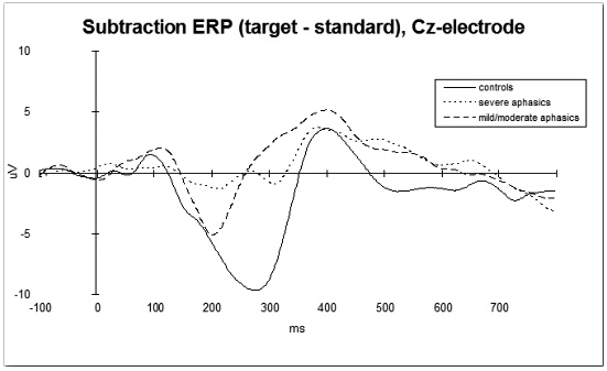
PHONEME PROCESSING IN APHASICS STUDIED WITH EVENT-RELATED BRAIN POTENTIALS IN A SYLLABLE DETECTION TASK
F. Becker, I. Reinvang (Nesoddtangen, NORW)
In this study of Norwegian language comprehension, we examined the neurophysiology of detecting infrequent target syllables (/ta/) amongst frequent standard syllables (/ba/). Event-related brain potentials (ERPs) were recorded from three groups: healthy persons (n=11), patients with severe aphasia (n=10) and patients with mild or moderate aphasia (n=9). The aphasics were grouped by Token test score (</>16.5).
All persons were able to detect the target syllables, but reaction time differed significantly: 383, 451, and 565 ms for controls, mild/moderate and severe aphasics respectively.
Three ERP components were of special interest: N1, P3, and N2, where N1 reflects processing of the standard syllable /ba/, P3 reflects processes involved in detecting the target /ta/, and N2 is the processing difference resolving from the subtraction of the ERP of the standard syllable from the ERP of the target syllable.
Analysis reveals differences between groups regarding all these components. With respect to the N1 component, there are no differences in latency, but in amplitude with the severe aphasics showing the smallest and the controls showing the largest amplitude. In addition, there are some hemisphere differences in the aphasics (reduced amplitude left vs. right) which are not found in the controls. A pattern similar to the N1 results can be observed for the P3 component.
The largest differences between groups are found regarding the N2-component: While the healthy subjects show a large negativity which can be recorded over the whole scalp and which extends from about 100 to 350 ms, the severe aphasics only show a small negativity. This difference seems to result from the lack of a central-parietal negativity with a peak at about 270 ms after the target stimulus in the severe aphasic patients. In addition, there are greater differences in amplitude between left and right hemisphere electrodes, which can not be observed in the controls. The mild/moderate aphasics hold an intermediate position. Interestingly, ERP latencies do not correspond with the reaction times recorded.
The results of this study will be discussed in the light of theories of auditory cognition and language comprehension models. This study suggests that ERP-measures – especially the negativity in the subtraction ERP – are correlated with the severity of language comprehension impairment.
At present, some aphasics are retested after rehabilitation; some first results will be presented as well.
O14 LOCOMOTION | C
WALKING-RELATED GAINS OVER THE FIRST 12 WEEKS OF REHABILITATION FOR INCOMPLETE TRAUMATIC SPINAL CORD INJURY: THE SCILT RANDOMIZED CLINICAL TRIAL
B. Dobkin, D. Apple, H. Barbeau, A. Behrman, D. Deforge, J. Ditunno, M. Saulino, R. Elashoff, S. Harkema, L. Fugate, M. Basso, M. Scott, G. Dudley (Los Angeles, USA)
Background: The Spinal Cord Injury Locomotor Trial (SCILT) was designed to compare two strategies for training patients with incomplete SCI to walk over the first 12 weeks of rehabilitation. The primary endpoint was 6 months after SCI. No studies have reported walking-related outcomes during rehabilitation for traumatic SCI.
Methods: A single-blinded, randomized trial conducted by 6 regional American and Canadian centers entered 107 ASIA C and D subjects and 38 ASIA B subjects who were unable to walk without physical assistance on admission for rehabilitation within 8 weeks of SCI. Subjects received conventional physical therapy (CONV) for mobility compared to the same amount of therapy employing body weight-supported treadmill training (BWSTT) and over ground practice. The Functional Independence Measure (FIM) for walking, walking speed for 50 feet, walking distance in 6 minutes, and lower extremity motor score (LEMS) were collected every 2 weeks during the intervention.
Results: Planned secondary analyses at 12 weeks revealed no significant differences in the ability to walk for UMN ASIA B and C subjects (p=0.41). ASIA B subjects were unable to walk. For ASIA C and D subjects, no differences were found between BWSTT vs CONV for walking speed (mean speed 51 vs 56 m/min, p=0.56) or walking distance. The LEMS correlated with walking speed (r=0.57) at 12 weeks, and 12-week walking speeds correlated with those at the primary endpoint at 6 months (r=0.91).
Conclusions: BWSTT and CONV therapy produced equal outcomes for walking parameters over the first 12 weeks of rehabilitation in ASIA B, C and D subjects. Most ASIA C and D subjects made remarkable improvements that were apparent by 5 weeks after starting inpatient rehabilitation. This data may help in the design of future clinical trials.
O15 FUNCTIONAL IMAGING | C
CEREBRAL PERFUSION CHANGES DETECTED IN MOTOR AREAS AFTER CONSTRAINT-INDUCED MOVEMENT THERAPY
I. Tarkka, M. Kononen, M. Husso-Saastamoinen, E. Vanninen, J. Kuikka (Kuopio, FIN)
Permanent hemiparesis is the most common deficit after stroke. Constraint-induced movement therapy (CIMT) belongs to a family of neurorehabilitation methods which emphasizes task-relevant repetitive training. Cerebral perfusion in twelve chronic stroke patients (mean duration of the disease 2.8 yrs; mean age 48 yrs) was studied to assess the effect of the two-week long CIMT rehabilitation. For SPECT imaging 550 MBq of Tc-99m-ethylcysteine dimer was intravenously injected similarly before and after the therapy. The SPECT scan was carried out 45-60 minutes later while the patient was resting. Mean total duration of supervised practice was 53 hours. A 16-task structured motor test was performed to assess changes in voluntary motor behavior. The mean total task time of the motor test decreased from 3.8 min to 2.5 min and functionality of movement scores improved significantly after rehabilitation. Images of those patients whose lesion was in the left hemisphere, were mirrored (i.e. all lesions were in the right hemisphere). Increased perfusion was found in motor control related areas. The specific areas with perfusion increase in the affected hemisphere were in precentral gyrus, premotor cortex (BA6) and in the frontal cortex, in superior frontal gyrus (BA10). In the non-affected hemisphere, perfusion was increased in superior frontal gyrus (BA6) and in cingulate gyrus (BA31). In the cerebellum, there was increased perfusion bilaterally. Perfusion decreased in lingual gyrus (BA18) in the affected hemisphere. In the non-affected frontal cortex, two areas with decreased perfusion were found in the middle frontal gyrus (BA8/10). Also the fusiform gyrus (BA20) and inferior temporal gyrus (BA37) showed decreased perfusion. Intensive therapy appears to facilitate motor restoration even in very chronic stroke patients who retain some residual motor control. Both the restoration seen in motor performance and in the changes of cortical perfusion at rest in motor related areas suggest that plastic processes can be enhanced by intervention also in the chronic state.
O16 LOCOMOTION | BS
INTER-LIMB COORDINATION CONTRIBUTE TO AMPLIFY LOCOMOTIVE NEURAL OUTPUT IN SPINAL CORD INJURED PERSONS
N. Kawashima, D. Nozaki, M. O Abe, M. Akai, K. Nakazawa (Tokorozawa Saitama, J)
It is well recognized that coordinated muscular activity can be induced by imposing locomotion-like movements even in the paralyzed lower limb muscles of complete spinal cord injured (SCI) persons. While the significant role of the afferent input related to hip joint movement and body load has been emphasized considerably in previous studies, it is still not fully understood to what extent the inter-limb neuronal pathway contribute to the human locomotive motor output. In the present study, we examine the effect of modalities of the contralateral leg motion on the magnitude of “locomotor-like” muscle activity.
The knee-locked leg swing movement was imposed on ten complete SCI subjects using a gait training apparatus. The following three different experimental conditions were adopted: (i) bilateral alternate leg movement, (ii) unilateral leg movement, and (iii) bilateral synchronous (in-phase) leg movement. In all experimental conditions, the passive leg movement induced the electromyographic (EMG) activity in the soleus muscle in all SCI subjects and in the biceps femoris muscle in eight of ten SCI subjects. On the other hand, the tibialis anterior and rectus femoris muscle didn’t show common synchronized EMG activity among the subjects. The EMG level quantified by integrating the rectified EMG activity recorded from the right leg was significantly larger for bilateral alternate leg movement than for unilateral and bilateral synchronous movements, although the right hip and ankle joint movements were identical in all experimental conditions. Further, the relation between EMG activity and applied load on the foot could not explain our results.
The present results suggest that the sensory information came from contralateral limb plays a substantial role in amplifying the induced locomotor-like muscle activity. It is reasonable to suppose that there is neuronal circuits enabling inter-limb coordination within the spinal cord, and this might contribute to regulate and shape human locomotive motor output.
O17 LOCOMOTION | BS
STEPPING-LIKE MOVEMENTS IN HUMANS WITH COMPLETE SPINAL CORD INJURY INDUCED BY EPIDURAL STIMULATION OF THE LUMBAR CORD: ELECTROMYOGRAPHIC STUDY OF COMPOUND MUSCLE ACTION POTENTIALS
M.M. Pinter, K. Minassian, F. Rattay, H. Binder, B. Freundl, F. Gerstenbrand, B. Freundl, M.R. Dimitrijevic (Vienna, A; Houston, USA)
Study Design: It has been previously demonstrated that sustained non-patterned electric stimulation of the posterior lumbar spinal cord from the epidural space can induce locomotor-like movements in subjects with longstanding complete spinal cord injury. In the present paper we explore physiologically related components of electromyographic (EMG) recordings during the induced stepping-like activity.
Objectives: To examine underlying mechanisms activated by electrical epidural stimulation of posterior lumbar cord structures effective to elicit stepping-like movements.
Material and Methods: The study is based on the assessment of epidural stimulation to control spasticity by simultaneous recordings of the electromyographic activity of quadriceps, hamstrings, tibialis anterior and triceps surae. We examined induced muscle responses to frequencies of 2.2–50 Hz in ten subjects classified as having a motor complete spinal cord injury (ASIA A and B). We evaluated stimulus-triggered time windows of 50 ms length of the original EMG traces. Stimulus-evoked compound muscle action potentials (CMAPs) were analyzed with reference to latency, amplitude, and shape.
Results: Epidural stimulation of the posterior lumbosacral cord recruited lower limb muscles in a segmental-selective way, which was characteristic for posterior root stimulation. 2.2 Hz-stimulation elicited stimulus-coupled CMAPs of short latency which were approximately half that of phasic stretch reflex times of the respective muscle groups. Their EMG-amplitudes were governed by the stimulus strength. Stimulation at 5–15 and 25–50 Hz elicited sustained tonic and rhythmic activity, respectively, and initiated lower limb extension or stepping-like movements representing different levels of muscle synergies. All EMG responses, even during burst-style phases were composed of separate stimulus-triggered CMAPs with characteristic amplitude modulations. During burst-style phases, a significant increase of CMAP latencies by about 10 ms was observed.
Conclusion: The muscle activity evoked by epidural lumbar cord stimulation as described in the present study was initiated within the posterior roots. These Posterior Roots Muscle Reflex Responses (PRMRRs) to 2.2 Hz stimulation were routed through monosynaptic pathways. Sustained stimulation at 5-50 Hz engaged central spinal PRMRR components. We propose that repeated volleys delivered to the lumbar cord can effectively modify the central state.
O18 MOTOR REHABILITATION | C
USAGE OF A COMBINED METHOD OF DYNAMIC PROPRIOCORRECTION AND PROGRAMMED ELECTRO-STIMULATION IN REHABILITAION AMONG NEUROLOGY PROFILE PATIENTS
K.V. Lyadov, I.V. Sidyakina, S.L. Zuev, T.V. Baidova, T.V. Shishova (Moscow, RU)
In our research we estimated the therapeutic effectiveness of a combined influence of a device “Gravistat” and a programmed electro stimulation using a computerized complex “Multimiostim” on the dynamics of movement status figures. An objective control-diagnosis criterion of the result of patients’ conditions was stabilometer scale – a method of estimation of patients’ vertical pose balance. Dynamic propriocorrection took place using “Gravistat”. The principle of the device lies in the effect of a strong flow of proprietor impulse on brain structures. It becomes a base for development and recreation of a misbalanced movement, patients’ emotional and will sphere. The flow is formed while the patient performs some movements wearing the “Gravistat” costume, which allows to set an individually dosed task, regulate it during the course of the recreation treatment. Programmed multi-channel electro-stimulation models physiological movement patterns, regulates person’s pathological movement stereotype, helps people with neuromotoric problems. Computerized complex “Multiomiostim” is responsible for the phase muscle micro stimulation while patient’s walking based on independence and synchronization.67 patients were under our research with pareses of different etymology: 43% – strong brain blood circulation misbalance, 34% – brain injuries, 15% – a spontaneous injury and neurosurgical operations, 8% due to pathology in spine brain. The research lasted from 3 weeks to 4,5 months. The first group of patients (36 people) in addition to their base treatment received treatments in costumes and multi-channel stimulation course, the other group – a standard course of recreation treatment. In the comparative analyses of the effectiveness of the patients’ rehabilitation in the two groups the following results were received: in the first group the figures of the statokinethiogram improved by 16,7% ( in the other group by 5,8%); the average speed of pressure center movement decreased by 19,1% (in the other group by 7,3%), the average square of the statokinethiogram decreased by 21,3% (9,8% in the other group ). The front pressure center place among the first group patients had an average of 12,2mm dynamics to the center, while 4,2mm in the second group. The shift of the pressure center in the sagittal part among first group patients had an average of 3,1mm, while 1,1mm among the second group patients. Thus, using a combined treatment of programmed multi-channel myo-stimulation and dynamic propriocorrection allowed to achieve better results in a shorter period of time, that proves the advantage of this method for recreation treatment among patients who suffer movement misbalance.
O19 BS
DIFFERENTIAL TIME COURSE OF NEUROPLASTIC PROCESSES FOLLOWING EXPERIMENTAL SPINAL CORD INJURY
U.H. Wiese (Cottbus, D)
Purpose: To test if patients with minimal volitional movements of the upper limb can regain better upper limb control and hand function using self-administered FES system.
Method: Only patients with ischemic stroke who despite intensive rehabilitation showed very little or no improvement 6 weeks after stroke onset (Fugl-Meyer [F-M] upper limb <10 at admission to acute rehabilitation, and <17, at 6 weeks post stroke) are considered in this report. Using randomized controlled trial, group 1 receive FES program (HandMaster system) plus standardized physical rehabilitation while group 2 receive standardized rehabilitation alone for 12 weeks. Training begins in the acute rehabilitation and continue at the patients’ residence. All patients practice 30 min, twice every day. Group 1 combine exercises with up to 4 hrs of daily FES to the forearm/wrist hand flexors/extensors using the Handmaster system. The stimulation is combined with task-specific training tailored and modified to each patient’s ability. Outcome measures of hand function (Box & Blocks [BB]; Jebsen-Taylor [J-T], and motor control [F-M] are recorded in addition to baseline at 4, 8 and 12 weeks.
Results: To date, 3 control and 3 experimental patients all with very slow recovery completed the trial. One FES treated patient recovered some hand function (able to perform the BB and J-T timed sub-test) and all 3 patients improved the F-M score considerably higher than the control patients who only gain few points on the F-M score.
Conclusion: The trend of faster and observationally greater recovery of motor control and better chance for re-learning hand function in these few patients suggest that as the study sample size increase these improvement should become both statistically and clinically significant. Based on previous data published by our group and other researchers worldwide, early initiation of FES training is clearly indicated.
O20 MOTOR REHABILITATION | C
BOTULINUM TOXIN IN THE TREATMENT OF SPASTICITY OF THE UPPER LIMB: A SYSTEMATIC REVIEW OF THE LITERATURE
T. Sycha, K. Elwischger, E. Auff, E. Fertl (Vienna, A)
Background: Botulinum toxin (BTX) has been used for the management of focal spasticity since several years, but data on treatment outcomes in upper limb spasticity (ULS) are scarce.
Objectives: With this systematic review we set out to clarify functional outcomes of BTX treatment (versus systemic myotonolytic agents, functional therapy, placebo, or no treatment) in ULS following stroke, traumatic brain injury or multiple sclerosis.
Methods: We searched the bibliographic databases MEDLINE, EMBASE, and the Cochrane Library from 1985 up to May 2004. We also reviewed the reference lists from identified articles and reviews of treatment studies. Furthermore we searched booklets of scientific congresses in the field of neurology, and contacted experts. Randomised controlled trials (RCTs) testing any dose of BTX for ULS caused by stroke, traumatic brain injury or multiple sclerosis, and describing functional improvement and subjective pain assessment were included in this review. All trials were quality scored and two independent reviewers extracted data. Results were compared for differences and discrepancies were resolved by discussion.
Results: Within 112 relevant citations we identified ten RCTs dating from 1996 to 2004. Nine RCTs reported on BTX type A, including 413 patients suffering from chronic post-stroke ULS and 37 patients with other reasons for chronic ULS. There was a single RCT testing BTX type B in chronic post-stroke spasticity of the upper limb. No RCT addressed the question of early versus late initiation of BTX treatment after stroke. Nine RCTs used placebo as control condition was. Injection sites were flexor muscles of the elbow, wrist and fingers. Pre-post modified Ashworth scores were measured in all studies. Pain ratings were reported in 3/10 RCTs. Various functional motor outcomes were measured in 6/10 RCTs. Subjective disability ratings were measured in 8/10 studies, and also carer burden was addressed in one trial.
The statistical combination of the study results (meta-analysis) is ongoing.
Reviewers’ conclusions: There is substantial scientific evidence for the efficacy of BTX-A in the reduction of chronic ULS caused by stroke. Treatment goals vary considerably between the RCTs and the focus on function and pain has just evolved during the last four years. There is still more work to be done in terms of optimal timing of BTX treatment after stroke, and in ULS caused by other cerebral disorders.
O21 REGENERATION | BS
AXONAL SPROUTING IN THE DENTATE GYRUS OF CAP23-OVEREXPRESSING MICE AFTER ENTORHINAL CORTEX LESION
D. Del Turco, G. Burbach, C. Gebhardt, A.G. Woods, J.P. Kapfhammer, M. Frotscher, P. Caroni, T. Deller (Frankfurt/Main, Freiburg, D; Basel, CH)
After entorhinal cortex lesion (ECL) the outer molecular layer (OML) of the dentate gyrus is denervated. In response to this deafferentation several fiber systems sprout and reinnervate the OML. However, only few commissural/associational (C/A) fibers invade the denervated area from the adjacent non-denervated inner molecular layer (IML). In order to study whether the sprouting response of C/A fibers can be enhanced by overexpression of the growth-associated protein cortical cytoskeleton-associated protein 23 (CAP23), ECL was performed in mice overexpressing CAP23 in adult neurons. The expression of CAP23 mRNA as well as the axonal sprouting response was compared in control and CAP23 transgenic (CAP23tg) mice. Non-radioactive in situ hybridization demonstrated that the sprouting neuron population expresses the transgene. Laser microdissection in combination with qRT-PCR revealed a very high level of transgene expression in the area of origin of the sprouting neurons. Anterograde tracing as well as immunocytochemistry were employed to analyze the sprouting response at the light- and electron microscopic level. In comparison to controls, CAP23tg mice showed an increased axonal sprouting response after ECL: Whereas only some C/A fibers invaded the denervated OML in controls, an enhanced invasion of the OML was observed in CAP23tg animals. In addition, sprouting fibers were longer in transgenic mice. These data indicate that CAP23 expression determines the extent of the C/A sprouting response in the hippocampus after ECL. (Supported by the DFG).
O22 PHARMACOTHERAPY | BS
THE VALUE OF DRUG TREATMENT IN THE FUNCTIONAL MOTOR RECOVERY AFTER STROKE: A CRITICAL REVIEW
J. Nijlant, M. Ijzerman (Enschede, NL)
Background: Different techniques are used to improve functional motor recovery after stroke. These vary between conventional physical therapy to more complex therapeutic regimens. Many of them have shown to be effective as far as motor control is concerned.
Another approach to be considered in the rehabilitation after stroke is the use of pharmacotherapy. In human some clinical trials have been carried out. Glycerol, selegiline, amphetamine and fluoxetine were used in different studies. Glycerol is a hyperosmolar agent which improves cerebral blood flow by reducing edema around cerebral infarcts. Selegiline is a selective irreversible inhibitor of monoamine oxidase type B and has both neuroprotective and neuronal rescuing properties. Amphetamine has dopaminergic, noradrenergic and serotonergic effects. Fluoxetine, a selective serotonin reuptake inhibitor, leads to an increased concentration of serotonin in the synaptic cleft. It influences brain processes.
The objective of the present review was to assess the available evidence about the effect of pharmacological modification of functional motor recovery after stroke
Methods: A systematic literature search was performed to identify randomised placebo controlled trials (RCTs) that have studied the effect of these drugs. The methodological quality of the studies was assessed by two independent raters. In particular outcome measures addressing motor control and functional gain were explored to judge the effect of drugs.
Results: Seven RCTs, with 428 patients in total, were included for this review. Three studies used amphetamine, two used fluoxetine, two used glycerol and one study used seligiline. The methodological scores ranged from 12–19. Commonly used functional outcome measures were Barthel index (5 studies) and the Fugl-Meyer assessment scale (4 studies). Of seven studies, four studies reported a positive effect: two studies of amphetamine (only when paired with physical therapy) and two studies of fluoxetine. For selegiline there’s no significant but some clinical motor improvement. Glycerol reduces mortality rates, but has no long term effect on functional motor recovery.
Conclusions: This review suggests a positive effect of amphetamine (only paired with physical therapy) and fluoxetine on the functional motor recovery after stroke. No firm conclusions can be drawn regarding the clinical implications of prescribing medication to improve functional motor recovery after stroke.
O23 PLASTICITY | C
VISION RESTORATION THERAPY (VRT) AFTER BRAIN DAMAGE: THE ROLE OF EYE MOVEMENTS AND FIXATION BEHAVIOR
B.A. Sabel, S. Kenkel, E. Kasten, I. Müller (Magdeburg, D)
Objective: We wished to study if eye movements and/or fixation are altered after vision restoration therapy.
Background: Patients with visual field deficits following stroke or neurotrauma can train their residual vision with VRT (Kasten et al., Nature med., 1998; www.novavision.info). VF-size increases have been demonstrated using super-threshold or near-threshold perimetry, but not with laser-scanning ophthalmoscopy (SLO; Reinhard et al., in press).
Methods: Data were compared among different, independent studies with respect to eye movements, border position shifts and fixation behavior, including determination of the blind-spot position.
Results: SLO measurements did not reveal evidence for eye movements before and after VRT greater than 1–2 degrees (Reinhard et al., unpublished). Only 1/3 of the patients showed slightly increased eye movements, but they were in the normal 1–2 range; in other cases, they remained unchanged. When analyzing the perimetric topography of the visual border position before and after VRT, border shifts were typically non-uniform: while in some areas VF border shifts clearly occured, in other areas they did not. Also, the shift of the visual field border in most patients was much greater than the expected 1–2 caused by normal eye movements (max. of up to 30 ). Furthermore, when different perimetry measures are compared, the border shift are not identical: while in some patients the border shift may be pronounced when a simple task is used (such as super-threshold perimetry), no border shift occurs when a difficult task is used (such as the SLO). Both the blind spot position and fixation quality measurements collected with standard perimetry remained unchanged before and after VRT.
Conclusion: VRT does not affect eye movements nor fixation in any significant way; rather true restoration of vision is possible.
O24 MULTIPLE SCLEROSIS | C
NEUROGENIC BLADDER DYSFUNCTION IN WOMEN WITH MULTIPLE SCLEROSIS-DIAGNOSIS AND PRIMARY TREATMENT OUTCOME
B. Schurch, A. Reitz, P. A. Knapp, S. Boy, A. Haferkamp (Zurich, CH; Heidelberg, D)
33 women with bladder dysfunction due to multiple sclerosis underwent clinical, neurological and detailed urodynamic examination and were treated according to complains and urodynamic results. Prevalence of clinical symptoms, urodynamic findings and primary treatment outcomes were analyzed.
Leading clinical symptoms were urgency in 87.9% followed by nocturia in 78.8% and urge incontinence in 72.7%. Most common urodynamic abnormalities were neurogenic detrusor overactivity in 63.6% and positive ice water test in 60.6%. In a group of 29 women who were able to void, incomplete voiding occurred in 79.3%. Anticholinergic medication was used in 26 patients with overactive detrusor, but 23% of the patients stopped medication after six weeks because of side effects like dry mouth, constipation and blurred vision. The alpha blocking agent tamsulosin was given to 18 patients with 100 ml residual urine and more. Although the drug slightly reduced residual volume, it was discontinued by 50% of the patients after six weeks because of side effects.
In conclusion, proper diagnosis and effective, well-tolerated treatment of bladder dysfunction in women with multiple sclerosis are challenging. A full urodynamic examination is recommended and may help to identify the underlying dysfunction. Troublesome side effects of the anticholinergic and alpha blocking medication limit patient compliance and lead to high attrition rates.
*BS – Basic Science; C – Clinical
© Hippocampus Verlag 2004
--------------------------------------------------------------------------------
|
